- PRODUCTSOverviewAbout our products range
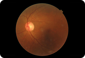
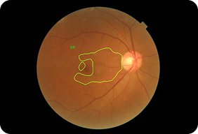
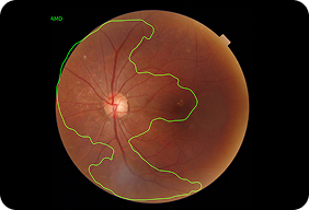
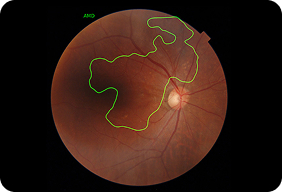
Dr.Noon is our AI retinal screening platform. Using a routine fundus image, it delivers dual insights: eye screening for key retinal findings and an instant cardiovascular risk assessment.
No new clinical process, no heavy infrastructure, just actionable screening insight.
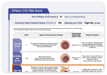
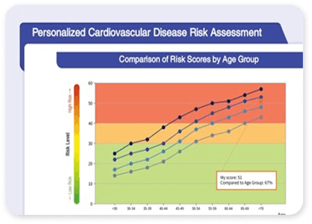
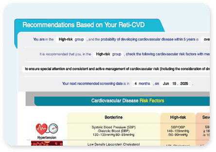
ProductsHeartInstant cardiovascular risk insight, clear risk banding output. Supports fast triage, prevention conversations, and timely referral.
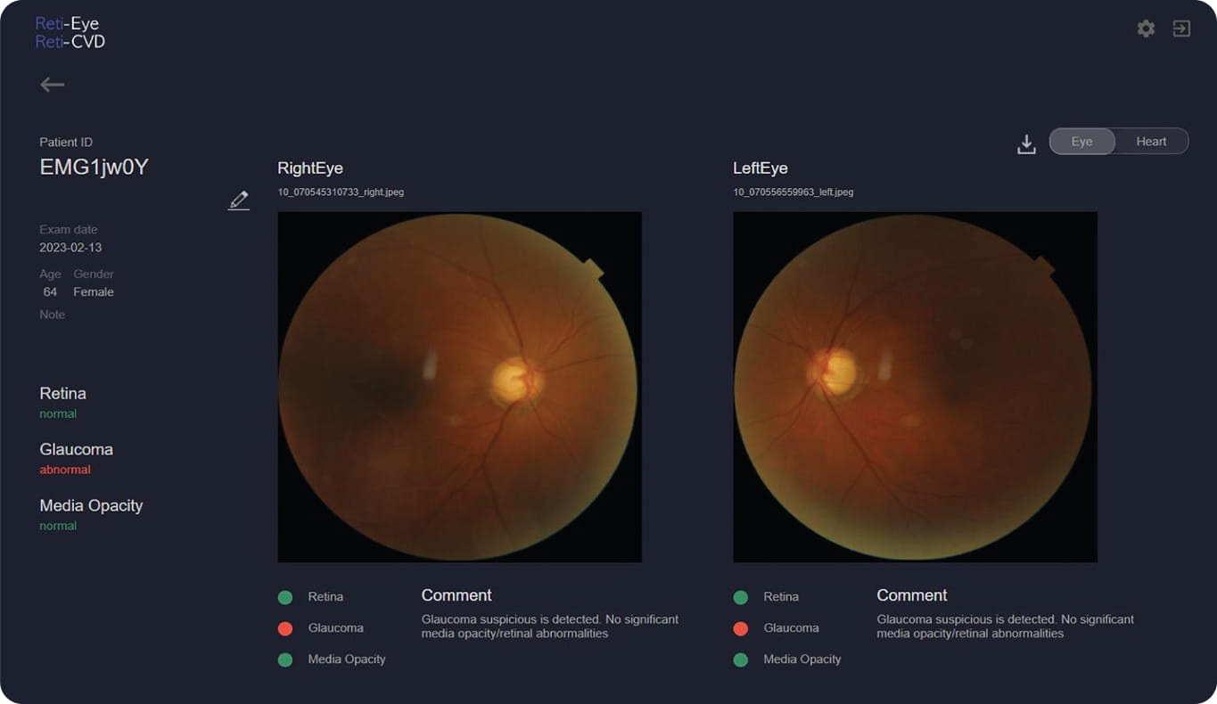
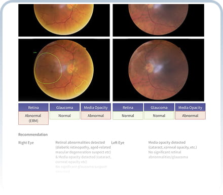
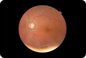
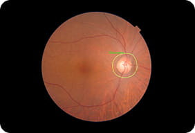
EyeAI retinal screening from routine fundus images, clear visual findings output. Confident clinical review, and timely onward referral.
ProfessionalsHealthcare ProfessionalsMinimal setup, flexible deployment model. Scale across clinics with consistent screening at speed.
Eye Care ProfessionalsAdds meaningful clinical value. Strengthen patient trust, differentiate your practice, and drive repeat visits.
- RESOURCESOverviewExploring Resources
Everything you need to support adoption and day-to-day use of Dr.Noon—clinical explainers, patient-friendly materials, FAQs, and evidence. Find quick answers, shareable resources, and practical guidance for confident conversations and smooth workflow integration.
ProductsResourcesBrowse our full library of guides, training materials, and practical tools to help you launch, run, and scale Dr.Noon in real-world settings.
PatientSimple, shareable explanations to help patients understand what the scan shows, what their results mean, and what to do next.
FAQsClear answers on workflow, image quality, reporting, referrals, and implementation, so your team can move faster with confidence.
DownloadsExplore the evidence behind Dr.Noon, including validation and research, ideal for clinical review, governance, and stakeholder confidence.
- CONTACTOverviewConnect with our team
Have a question about Dr.Noon, onboarding, compatibility, or rollout? Our team can help with demos, implementation planning, and practical guidance so you can start screening quickly and confidently. Get in touch and we’ll point you to the right next step.
Ways to get in touchReady to bring Dr. Noon into your practice?